
7.2.1. White line disease (onychomycosis) associated with abscess and hoof bone rotation
2. Data of the patient
Breed: German Warmblood
Sex: mare
Age: 17 years
Color: dark brown
Working discipline: breeding, hobby
3. Anamnesis
-
Reason why the owner complains: Damaged white line and loose hoof wall. Significant lameness in an extensive abscess in the dorsal part of the hoof.
-
Duration of the problems: 2,5 years
-
Stabling conditions: Box, paddock.
-
Bedding: straw
-
Frequency of the hoof care: Every 6 - 8 weeks
-
Type of shoeing: Originally without shoes, after onychomycosis, shod with quarter clip horseshoes.
-
Lameness and diagnosis: Significant lameness on the left forelimb.
4. Case description
-
Characterization of the problems: When trimming the left front hoof, a damaged white line and a separation of the hoof wall from the foot were observed. Due to the infection, which penetrated the damaged white line on the hoof wall, a large abscess formed on the inner front part of the hoof wall and significantly worsened the lameness.
-
Hoof shape and pathological changes: The veterinarian removed the hoof wall from the affected part of the hoof and opened the abscess.
-
Conformation: Limb posture had no effect on the disease.
-
Evaluation of the hoof care: No method of treatment was recorded before the procedure.
-
Evaluation of the shoeing type: The hoof was not shod.
5. Chosen solutions
-
Trimming: During the trim, the heels were lowered to achieve a better anteroposterior balance of the hoof.
-
Shoe preparation: The front quarter clip horseshoe Mustad size 2, size 22x8 mm was chosen for shoeing.
-
Shoeing: To support and protect the foot, the hoof was shod with a leather pad with a Luwex 120 soft hoof packing , see Fig. No. 1 - 3. of 17.8.

Fig. no. 1.

Fig. No. 2.

Fig. No. 3.
Veterinary treatment: Occasional disinfection and check up of the transition between the growing hoof wall and the horn covered coffin bone laminar corium.
Rules of the further care:
Regular shoeing at one-month intervals with check-up of the correct growth of the hoof wall
6. Follow up – Development of changes
Effect of the first trim:
The hoof was stabilized with aquarter clip horseshoe and foot protection. Also, better anteroposterior balance reduced the pain in the hoof and improved movement of the horse. In this way, the hoof was shod five more times at regular monthly intervals.


Fig. No. 4., 5., 6. - 2. shoeing 20.9.2017


Fig. No. 7., 8., 9. - 3. shoeing 19.10.2017


Fig. No. 10., 11., 12. - 4. shoeing 7.12.2017


Fig. No. 10., 11., 12. - 5. shoeing 16.1.2018

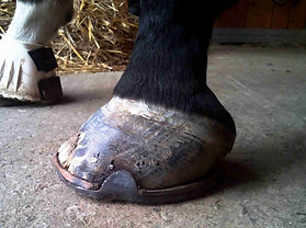
Fig. No. 13., 14., 15. - 6. shoeing 5.2.2018
Gradually, however, according to the direction of the coronary band, a probably wrong direction of the hoof wall was found. Also, its quality was not good, especially on the inner side of the hoof, which indicated a continuing disease of the white line, see. Fig. No. 16 and 17.

Fig. No. 16. - Continued onychomycosis

Fig. No. 17. - Wrong direction of the hoof wall growth

Changes in shoe choice and shoeing:
Before further shoeing on March 15, 2018, the hoof was x-rayed. The image showed a rotation of the hoof bone of 21 °, high heels and a bruised sole under the tip of the hoof bone, see Fig. No. 18.
The hoof was trimmed according to the rules for laminitic hooves with respect to the outcomes of the X-ray image. The dorsal wall was rasped parallel to the dorsal surface of the coffin bone. This procedure also removed a poor quality hoof wall up to a healthy horn, see Fig. No. 19., 20., 21.
Fig. No. 18. - X-ray image from March 15, 2018


Fig. No. 19., 20., 21 .: From left - Hoof after trim 15.3.2018


Fig. Nos. 22, 23, 24, 25, 26:
From left - Left front hoof after shoeing on March 15, 2018
The hoof was shod with a quarter clip front horseshoe Libero Equilibrium size 2 size 22 x 8. Because a large part of the hoof wall in the dorsal and medial part of the hoof was removed, fullers were extended to the ends of branches and two new nail holes were stamped on both sides of branches. A heart bar was welded between the ends of the branches to the level of the shoe foot surface so as to create active pressure on the frog and provide firm support to the rotated coffin bone.
Farriery treatment effect: - The hoof was shod three times in this way.


Fig. No. 27.,28.,29.,30.,31.,32. - Trim and shoeing on 3.5.2018


Fig. No. 33., 34., 35. - Trim and shoeing on 21.6.2018


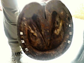
Fig. No. 36., 37., 38. - Trim and shoeing on 14.8.2018


Fig. 39., 40 .: X-ray check up
Seven weeks after the third shoeing with a heart-bar shoe, control X-rays were taken. The position of the coffin bone was improved and the palmar angle was reduced.
Hoof wall grew parallel with the dorsal surface of the coffin bone and its quality on the bearing edge of the hoof gradually improved. This allowed for a safer nailing of the horseshoe in the side parts of the bearing edge of the hoof.
Result of the care:
After 15 months of farrier care, the hoof was stabilized and the horse's movement was comfortable. After a few more shoeings, the hoof wall will grow to the sole, but the white line will probably remain damaged. It will require constant care and check ups, as it will continue to be an entrance for bacterial and fungal infections.


Fig. No. 41., 42 - Shoeing on 15.11.2018
The long-neglected white line disease - onychomycosis very often leads not only to hoof damage, but also to the loosening of the laminar connection and mechanical laminitis. Any white line defect must be addressed intensively, because it can very easily turn into long-term treatment with irreparable consequences.
8. Conclusion (take home message):