
9.4.1. Ossification of lateral cartilages
1. Author
Breed - Czech warmblood
Sex : mare
Age 13 years
Working discipline: former sports mare, currently a hobby due to long-term health problems
2. Data of the patient
Reason why the owner complains: Long-term lameness on both front limbs with a reaction to the load on a hard surface.
Duration of the problems: More than one year.
Stabling conditions: In the stall.
Bedding: straw
Surface on which the mare predominantly moves: Sandy riding arena, but terrain with a diverse surface predominates.
Frequency of the hoof care: Regularly every 7-8 weeks
Type of shoeing: At the time of the examination, the mare was barefoot.
3. Anamnesis
Characterization of the problems:
Alternating lameness on both front limbs lasting more than a year. The lameness gradually worsens and is more pronounced in turns on hard surfaces. The left limb is worse.
Conformation:
There are no significant conformation faults, only a slight offset carpus and inward rotation of the finger load the inner parts of the limbs and hooves.
Hoof shape and pathological changes - Shape of the hoof and pathologic changes:
The hooves are differently positioned in the sagittal plane, the left is sloping and the right is steeper. In the horizontal plane, the inward rotation of the pgalanges creates an internally diagonal hoof shape.
Evaluation of the hoof care:
Regular trims of the hooves and movement of the horse was the reason for the good condition of the horn in a reasonable length and the corresponding shape of the hooves.
Evaluation of the type of shoeing:
At the time of the examination, the mare was barefoot.
Examination results:
-
The mare was presented on a straight line and in both circles on a soft and hard surface. The most intense 3rd grade lameness was on the hard surface on the left circle on the inner limb.
-
Flexion tests did not significantly worsen the degree of lameness.
-
Local anesthesia on the feet was 80% positive on both limbs after 5 minutes.
-
Subsequent X-ray examination revealed ossification of lateral cartilage at grade 3-4.


Fig. No. 1-3: From left - lateromedial, dorsopalmar and oxspring X-ray of the right front
hooves. Medial cartilage on both limbs showed a greater degree of ossification.


Fig. No. 4.-6 .: From left - Lateromedial, dorsopalmar and oxspring X-ray of the left front
hooves. Medial cartilage on both limbs showed a greater degree of ossification.

4. Problem description
5. Chosen solutions
Selected hoof trim:
-
The heels were lowered on both hooves, especially on the right hoof, where the cartilages were compressed by the hoof capsule due to the high heels.
-
The bearing edge of both hooves was aligned parallel to the solear surface of the coffin bone, depending on the height of the arch on both halves of the hooves.
Shoe preparation and shoeing:
-
Horseshoes Libero - Equilibrium front two-clip size 2 with full-roll effect on the sides were chosen for shoeing. The shoes were fitted only with small overlaps with the intention of preventing their roll effect.
-
Luwex senior pads were used in the shoeing in combination with the M9 hoof packing material, in order to dampen vibrations during impact of the hooves and to partially reduce the hoof mechanism in the palmar parts of the hooves.


Fig. No. 7.-8 .: From the left - Right and left front hoof when viewed from below.
Veterinary treatment: If necessary to relieve pain and inflammation, one sachet of Danilon per day.
Rules of the further care:
Regular adequate movement on a soft surface on straight lines, with absolute restriction of movement on a hard surface.
6. Follow up – Development of changes
Effect of the first selected hoof treatment - Trimming effect:
The first treatment of hooves and shoeing with a cushioning effect brought an improvement of about 50%. No further improvement in the degree of lameness was observed after the second shoeing in this way. Although the moisture under the pad supported the flexibility of the hoof capsule, it caused the development of the thrush of the frog, which was not as abd before the first shoeing. The hoof capsule in the area of ossified cartilage was too tight for the degenerated cartilage and the constant irritation of the joint was painful.
Changes in the choice of horseshoes and shoeing:
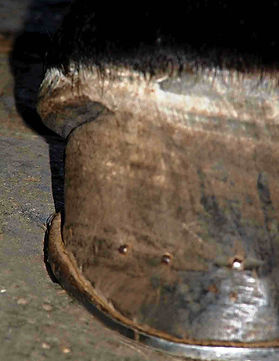
Further shoeing was performed on a single-clip keg shoe with a rocker and adequate overlaps of the branches. The complete Colin's release grooves were created in the quarter parts of the hooves using an angle grinder. The grooves were ground with a 4 mm thick grinding wheel through the entire thickness of the hoof wall almost almost up to the wall corium. The horizontal groove was made under the coronary band at the point of transition of the coronary cushion into the wall laminae. The horn under the coronary band was ground until a slight infiltration of blood. A thin layer of the horn then did not prevent the coronary band from expanding and growing out of the more spacious hoof capsule. Just before the horseshoe was nailed, a part of the bearing edge between the two vertical cuts was ground so that the bearing edge did not lie on the shoe and did not transmit pressure to the loose coronary band.


Fig. No. 9.-10 .: Complete Colin's release hoof groove. The grooves were ground with a 4 mm thick grinding wheel through the entire thickness of the hoof wall almost almost up to the wall corium. The horizontal groove was made under the coronary band at the point of transition of the coronary cushion into the wall laminae. The horn under the coronary band was ground until a slight infiltration of blood.


Fig. No. 11 .: Complete Colin's release hoof groove. Front view of the hoof before shoeing
Fig. No. 12 .: Just before nailing the horseshoe, a part of the bearing edge between the two vertical cuts was ground so that the bearing edge did not lie on the shoe and did not transmit pressure to the loose coronary band.


Fig. No. 13.-14 .: View of the trimmed and shod hooves from behind and from the side.
The effect of farriery measurest:
Already after the first shoeing interval, the widening of the coronary band in the lateral parts of the hooves was clearly visible. By this measure, the ossified cartilage gained almost 2 cm of space in the newly grown hoof capsule (see Fig. 15).
Result of the care
After the whole hoof capsule grew, the mare stopped limping completely on the soft surface . During long work on a hard surface, problems with limping occasionally returned, but to a much lesser extent. With reasonable management, she was able to perform the work of a hobby horse with occasional jumping over smaller obstacles.
Fig. No. 15 .: View of the extended growing coronary band
7. Conclusion (take home message):
Hoof cartilage ossification is a progressive and very difficult to treat disease of the palmar part of the hoof. Cartilage ossification usually takes place gradually at various lengths of intervals.
In the first stages, only active ossification centers cause pain, and after the acute phase calms down, the sensitivity of the hooves subsides. However, in the case of strongly ossified cartilage, proper shoeing and management usually no longer help. Then the only other option is a release hoof grooves.
The result of correctly made release cuts is twofold. The vertical cuts serve to quickly release the circumference of the hoof capsule in the side parts, but this effect is not very strong. From a long-term point of view, the horizontal part of the incision has the greatest positive effect, which is able to widen the hoof in the lateral and heel parts and thus provide a larger space for enlarged and inelastic cartilage. In many cases, however, even cuts do not help and the horse is doomed to persistent palmar pain.