Title (Diagnosis): 8.4.2. Laminitis
1. Author of texts and photographs: Ing. Jindrich Vincalek, CE-F
Place of work: Veterinary clinic Heřmanův Městec
2. Literary review: Horseshoeing, 42.6. Laminitis ,ISBN: 978-80-7490-052-5, Print Bat 2015
3. Patient data No. 8.4.2.
Breed: Czech Warmblood
Sex: Mare
Age: 6 years
Color: Dark Bay
Work use: Breeding and recreational riding
-
Problems complained of by the owner: Severe pain in the front hooves, degree of lameness 4-5 when moving, mare lying down, not willing to get up
-
Duration of the problem: 6 months
-
Stables: Box
-
Feed : Hay, core
-
Litter : Straw with shawings
-
Frequency of hoof treatment : Before the disease barefoot, shod after the first signs of laminitis, after symptoms disappearance again barefoot with a trim every 2 monhs. Last 14 days - plaster bandages at the clinic.
-
Type of shoeing: Barefoot
-
Lameness, possible diagnosis: Currently severe pain in the limbs on impact and in the weight bearing phase of limb movement.
4. Anamnesis
5.1. Characteristics of changes:
The mare has difficulty moving even in plaster bandages around a box bedded high in shawings. She lies often and for a long time and shows a reluctance to get up. There are signs of bedsores on the flanks and hips. The mare shows all signs of recurrence of laminitis.
5.2. Limbs conformation
The mare typically pushes the hind legs forward in an effort to relieve the load on the forelegs. The forelegs have a camped out stance, which partially reduces the pull of the deep digital flexor tendon on the coffin bone and transfers the load to the palmar part.
5.3. Hoof shape and pathological changes:
The shape of the hooves of the forelegs is typical of chronically laminitic hooves.

Fig. 1: Seen from the side, the dorsal wall of both hooves is strongly concavely curved. The heels are high and cause a double impact. The growth rings diverge from the dorsal part of the coronary band towards the heels and prove a slower hoof wall growth in the front part of the hoof, where the coffin bone connection is damaged due to its rotation and descent. From this point of view, it documents the change in the position of the coffin bone as well as the constriction of the wall just below the coronary band.
5. Case description


Fig. No. 2 .: Right front hoof
Fig. No. 3 .: Left front hoof
When looking at the hooves from the front, a slight divergence and constriction of the hoof capsule under the coronary band is visible. The coronary band is dropped in the middle of the dorsal part, which is an accompanying sign of a sinking of the hoof bone.


Fig. No. 4 .: Right front hoof
Fig. No. 5 .: Left front hoof
Looking at the soles, we confirm a significant growth of the horn and its excessive height. In palmar part of the frog and heel bulbs is visible significant damage caused by thrush.

Fig. No. 6 .: Sole of the right hoof

Fig. No. 7 .: Sole of the left hoof

Fig. No. 8 .: Sole of the left hoof
Figs. 6. and 7.:
The look on the soles of both hooves shows considerable dorsopalmal imbalance created by the rotation of the coffin bone and subsequent long-term poor trims. In front of the tips of the frogs are noticeable bruises created by the pressure of the tips of the sunken coffin bones. In the right hoof, the infiltration of blood into the horn is more pronounced due to the greater descent of the coffin bone and smaller thickness of the sole.
Fig. No. 8 .:
A stretched, about 15 mm wide white line can be seen in the dorsal parts of the bearing edges of both hooves as a result of the rotation of the coffin bones.
5.4 .: Evaluation of the trim:
The strongly neglected shape of the hooves, the discomfort when standing and moving testify to the long-term poor trimming and shoeing. A curved dorsal wall, poor dorsopalmal balance, high heels, poor breaover point and the thickness of the soles at the minimum limit are clear symptoms of unprofessional care for laminitic hooves.
5.5 .: Evaluation of previous shoeing:
The hooves were barefoot. On the right hoof was remainder of the plaster bandage, which was difficult to remove because the mare refused to stand on her left limb.


Fig. No. 9 and 10: X-rays before shoeing. By evaluating the images, a smaller drop of the coffin bone, greater rotation and a 3 mm thicker sole were found on the right hoof (Fig. No. 9 on the left) than on the left hoof (Fig. No. 10 on the right). This is the reason why the left hoof was more sensitive and painful.
6. Documentation - X-rays
7.1. Chosen trim: The principle of the laminitic hoof trim is in adapting the shape of the hoof capsule to the shape and position of the coffin bone.


Fig. No. 11 and 12 .: View of the left front hoof from the side and front.
On both hooves, the concavely curved dorsal wall was first shortened and leveled with hoof nippers and rasp so that it was parallel to the dorsal surface of the coffin bone.


Fig. No. 13 - 17.: View of the right front hoof from the side and from behind.
Then the sole was trimed with respect to keep its maximum thickness in front of the tip of the frog. In the dorsal part of the bearing edge, a long rocker was rasped to ensure breakover as close as possible to the tip of the coffin bone.


Fig. No. 18 and 19 .: From the left - view of the right and left foot after trim before shoeing
This treatment ensured a positive dorsopalmal balance of both hooves, which is very important for laminitic hooves.
7.2. Shoes preparation:
Equilibrium quarter clip shoes were chosen for shoeing, which allow the shoe to be set back and their forged rocker mproves the dorsopalmal balance of the hooves. During fitting, we bent the dorsal part of the shoe according to the rocker on the hooves, which maximally reduces the tension of the deep digital flexor tendon and its negative effect on the coffin bone. After fitting, "mercedes" spider plates were welded on the shoes to the level of the bearing surface, in order to increase the distance of the coffin bone tip from the surface. This type of bars provides the palmar part of the hoof and frog with sufficient and firm support.


Fig. No. 20 and 21: Fitted and modified shoes
7.3. Shoeing
A special hoof packing material for laminitic hooves - Luwex Premium - was used for the shoeing itself, which filled the collateral grooves and the sole in a thickness of about 1.5 cm. The bearing edge of the hoof must be left without packing. Just before the packing hardened, a shoe was nailed on the hoof with two nails and the limb was placed on the ground so that the packing was perfectly pressed between the horseshoe and the arch of the foot. The remaining nails were then hammered into the back holes of the shoe. The importance of a strong cushion lies in the protection of the weak sole under the tip of the coffin bone and in the stable support of the lower surface of the coffin bone against the drop and effect of rotation.


Fig. No. 22., 23., 24., 25 .. 26 ..:
Right front hoof after shoeing


Fig. No. 27, 28, 29, 30, 31:
Right front hoof after shoeing
7.4. Veterinary measures
X-ray examinaton at the next shoeing
7.5. Principles of further care
Secure shoeing at regular intervals according to the growth of the horn and the balance of the hoof. Stable mare in high layer of shavings with limited movement for 4 - 6 months. Feeding restrictions in order to keep the mare's weight at the lower limit.
7. Chosen measures
8. Development of changes
8.1. Effect of the 1st selected hoof adjustment
The effect of the hoof treatment was immediately visible. By a reasonable reduction of the heels, the large palmar angle of the coffin bones with respect to the bearing edge of the hooves was reduced, which significantly reduced the pressure of the coffin bone tip on the sole and thus the pain of the hooves.
8.2. Changes in the choice of shoes and shoeing
The choice of the trim and the method of shoeing proved to be appropriate. The chosen method of shoeing significantly reduced the pain of the hooves and increased the comfort of the mare's movement. It will not be necessary to change the shoeing method in the next shoeing intervals.
8.3. The effect of farriery measures
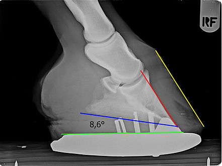
The modification of the hooves was focused mainly on improving the shape of the hoof capsule with respect to the position of the rotated coffin bone. By lowering the heels and leveling the concavely curved dorsal wall, a positive dorsopalmar balance and a better load on the lower surface of the cofin bone were achieved. The palmar angle of both coffin bones was reduced by 50 %. The Equilibrium shoe with a rocker allowed the breakover point to be moved up to the tip of the coffin bones of both hooves and limited the pull of the deep digital flexor tendons. The "mercedes" spider plate bar together with the Luwex Premium hoof packing created protection and at the same time support for the soles overloaded with rotated coffin bones.
8.4. The result of care
Before shoeing, the mare had difficulty moving around the deep-bedded box and could not walk down the concrete corridor to be shod. After shoeing, she was able to move relatively comfortably even on hard surfaces


Figs. No 32 and 33:
X-ray of the right front hoof before and after shoeing
Figs. No. 32 and 33:
X-ray of the left front hoof before and after shoeing

Schvácené kopyto
Proper trim of the hoof capsule is very important for laminitic hooves. It must ensure a good dorsopalmar balance, which affects the load on the coffin bone and eliminates the pull of the deep digital flexor tendon. The dorsal wall must be as light as possible so that the pressure does not prevent the new wall from growing off at the coronary band. The shoeing method must support the balance of the hoof while protecting the overloaded soles. Adherence to the principles of treatment and shoeing of the laminitic hooves gives the affected horses relative comfort when moving and a greater chance of recovery.

